Technology
Four tools, and the specific question each one answers.
A list of equipment is not a reason to choose a practice. What matters is what the machine changes about the decision — so each of these says what it is for, when we use it, and when we do not.
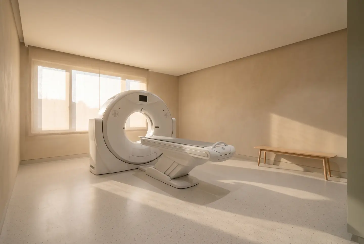
01
Cone beam CT
A three-dimensional radiographic scan of a defined volume of the jaw.
It is what makes guided implant surgery possible: bone height and width, bone density, the exact path of the inferior alveolar nerve and the floor of the sinus, measured rather than estimated. We plan the fixture against the crown we intend to make, print a surgical guide from that plan, and place through the guide.
- When we use it
- Every implant case. Some difficult endodontic and impacted-tooth assessments. Not routine screening.
- The caveat
- A single-arch CBCT delivers a higher dose than a panoramic radiograph and a much lower dose than a medical CT. We take the smallest field of view that answers the question.
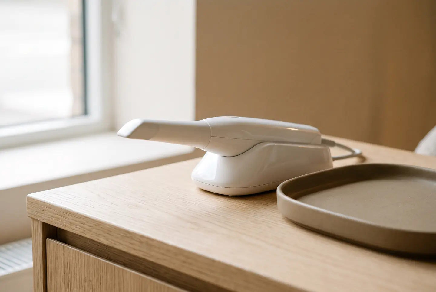
02
Intraoral digital scanning
A handheld wand that builds a three-dimensional model of the teeth from a video stream.
It replaces impression material for most work, which most patients care about more than the accuracy. It also means the model is a file: we can overlay this year on last year to measure wear, design a restoration against the opposing arch, and send the case to the laboratory the same afternoon.
- When we use it
- Aligners, veneers, crowns, night guards, implant restorations, and wear monitoring.
- The caveat
- No radiation. Some deep subgingival margins still scan better with a conventional impression, and we will say when.
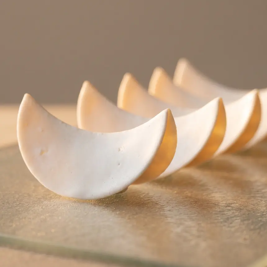
03
Digital smile design and trial smiles
The proposed shapes designed on your photographs and scan, then milled or printed as a physical mock-up.
A design argued about on a screen is not the same as a design worn to dinner. The mock-up is trialled directly on unprepared teeth in temporary material, so you can photograph it, sleep on it and come back with changes while everything is still reversible.
- When we use it
- Every veneer case and every full-mouth rehabilitation. Often for bonding too.
- The caveat
- The design also sets how much enamel has to be removed — preparation follows the approved shape rather than a fixed depth.
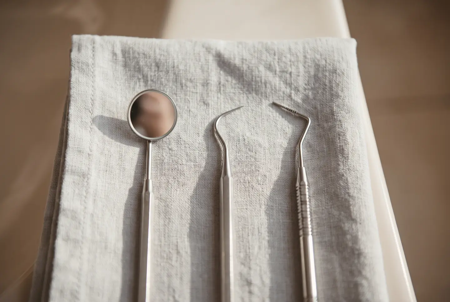
04
Photographic records
A standardised series at the first visit and at every review, shot to the same framing and lighting.
Standardised photographs are the only honest way to compare a mouth to itself. They are how we show a whitening result against a shade tab rather than against memory, and how we spot wear progressing before a patient notices it.
- When we use it
- First visit, before and after every cosmetic case, and annually thereafter.
- The caveat
- Your records stay yours. Nothing is published anywhere without separate written consent.
What we have not bought
Some of it is marketing.
We do not have a chairside milling unit. A milled crown in an hour is genuinely convenient, but for the anterior work this practice mostly does, a ceramist with a photograph and a stump-shade record produces a better result than a machine and a shade tab. So our ceramics go to a laboratory and you wear a temporary for two or three weeks.
We do not offer laser gum contouring as a standalone cosmetic service. Reshaping a gum margin is part of a designed case with a periodontal assessment behind it, not a twenty-minute add-on.
And we do not use whitening accelerator lights. The published evidence for them is weak; what produces the result is the peroxide, the concentration and the contact time.